Chapter 9
Pleura, Chest Wall, and Diaphragm
Learning Objectives
- Recognize and name four causes of a large unilateral pleural effusion on a chest radiograph or computed tomograph (CT).
- Describe the difference in appearance of a pulmonary abscess and an empyema on chest radiograph or CT.
- Recognize diffuse pleural thickening, as seen in fibrothorax, malignant mesothelioma, and pleural metastases.
- Recognize a pneumothorax on an upright and supine chest radiograph.
- Recognize imaging findings suggesting a tension pneumothorax or hydrothorax and describe the acute clinical implications.
- Recognize a pleural-based mass with bone destruction or infiltration of the chest wall on a chest radiograph or CT; name four likely causes.
- Recognize pleural calcification on a chest radiograph or CT and suggest the likely diagnosis of asbestos exposure (bilateral involvement) or old tuberculosis or trauma (unilateral involvement).
- Recognize the typical chest radiographic appearances of pleural effusion, given differences in patient positioning, and describe the role of the lateral decubitus view to evaluate pleural effusion.
- Recognize apparent unilateral elevation of the diaphragm on a chest radiograph and suggest a specific etiology with supportive history and associated chest radiograph findings.
The chest wall, pleura, and diaphragm enclose the outer
lung. All are intimately associated with each other, which occasionally
makes it difficult to determine the origin of a mass involving one or
more of these compartments (Fig. 9-1).
Disorders involving the chest wall, pleura, or diaphragm - resulting in a
"pleural-based" mass - can arise from one of these compartments, an
adjacent compartment, or another part of the body (as with metastatic
neoplasms). Certain radiologic features can help determine the origin
of an apparent pleural-based mass and narrow the list of diagnostic
possibilities.
Pleura
The pleura is composed of visceral and parietal serous
membranes. The lungs and interlobar fissures are invested in the
visceral pleura, whereas the parietal pleura lines the ribs, diaphragm,
and mediastinum. The visceral and parietal pleura are continuous with
one another as they are reflected around the hilum and the inferior
pulmonary ligament. Inferiorly, the parietal pleura is situated within
the costophrenic sulcus. The area between the two pleural layers forms
a "potential space," which can be enlarged when filled with fluid,
cells, or air. Normally, there is approximately 1 to 5 mL of pleural
fluid within this space (1). Because the total
thickness of the pleural space and the normal visceral and parietal
pleura is only 0.2 to 0.4 mm, the pleural layers are not usually
identified on radiographs or computed tomographic (CT) scans of the
chest except (a) when outlined by air or extrapleural fat, (b) where
the visceral pleura invaginates into the lung to form the fissures, or
(c) where the two lungs contact each other at junctional lines (2).
The parietal pleura is separated from the ribs and intercostal muscles
by a layer of fatty areolar connective tissue and a layer of
endothoracic fascia. The parietal pleura receives vascular supply from
and is drained by the systemic circulation, whereas the visceral pleura
is supplied and drained mainly by the pulmonary circulation. Lymphatic
drainage of the visceral pleura is by way of a lymphatic plexus that
covers the surface of the lung just beneath the visceral pleura (3).
These lymphatics do not connect with the pleural space. The parietal
pleura is the primary drainage route for fluid in the pleural space,
as it contains lymphatics that connect to the intercostal, internal mammary, and mediastinal lymph node chains (3). Except in rare circumstances (e.g., in some cases after cardiothoracic surgery or trauma), the right and left pleural spaces do not communicate with each other.
P.139
as it contains lymphatics that connect to the intercostal, internal mammary, and mediastinal lymph node chains (3). Except in rare circumstances (e.g., in some cases after cardiothoracic surgery or trauma), the right and left pleural spaces do not communicate with each other.
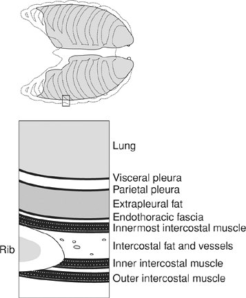 |
FIGURE 9-1. Association between the lung, pleura, and chest wall.
The lungs are invested in the visceral pleura, whereas the parietal
pleura lines the ribs and soft tissues of the chest wall, diaphragm,
and mediastinum. The parietal pleura is separated from the ribs and
intercostal muscles by fat and endothoracic fascia. The total thickness
of the pleura and "potential" pleural space is only 0.2 to 0.4 mm. |
A pleural-based density is one that, in some projection,
presents a more or less sharp border indicative of a pleural surface,
with a projected center lying outside the parenchyma of the lung. It
may be in one of five locations: (i) extrapleural, (ii) parietal
pleural, (iii) interpleural, (iv) visceral pleural, or (v) subpleural (Fig. 9-2).
When associated with a rib lesion, an extrapleural lesion is most
likely a hematoma (as from a rib fracture) or a tumor (with rib
metastasis); when there is no rib lesion, it is likely a lipoma or
lymphoma (when a patient is known to have lymphoma). Lesions involving
the parietal pleura are usually calcified and represent plaques from
prior asbestos exposure. Interpleural density is common and can
represent loculated pleural effusion or metastatic tumor. Visceral
pleural lesions are uncommon but usually represent pleural thickening
from asbestos exposure (whereas calcified asbestos-related plaques more
frequently involve the parietal pleura). Subpleural densities are
parenchymal and generally do
not have a sharp margin with the lung (Fig. 9-3), but on occasion tumors in the apex of the lung (so-called Pancoast tumors) can appear fairly well circumscribed (Fig. 9-4). The etiology of many soft tissue masses, regardless of their origin, cannot be distinguished on radiography or CT of the chest, except in the case of lipoma, which has a characteristic low attenuation value on CT.
P.140
not have a sharp margin with the lung (Fig. 9-3), but on occasion tumors in the apex of the lung (so-called Pancoast tumors) can appear fairly well circumscribed (Fig. 9-4). The etiology of many soft tissue masses, regardless of their origin, cannot be distinguished on radiography or CT of the chest, except in the case of lipoma, which has a characteristic low attenuation value on CT.
 |
FIGURE 9-2. Radiologic pleural-based densities.
A pleural-based density has margins that are partially or completely
well circumscribed, indicating contiguity with a pleural surface. There
are five types of pleural-based densities, depending on the location of
origin. An extrapleural densityoriginates
in the chest wall, and when it does not extend into the pleura and
lung, it has a sharp medial margin where it contacts the parietal
pleura. When the adjacent rib is involved, rib fracture with hematoma
or neoplasm should be considered. Parietaland visceral pleural densitiesare usually asbestos-related pleural plaques, which may or may not be calcified. Interpleural densitiesmost
commonly represent loculated pleural effusions; when intrafissural in
location they may be recognized on chest radiography by characteristic
"beaking" at the ends of the fluid collection where the pleural layers
of a fissure meet (producing a "pseudotumor"). Subpleural densitiesare
parenchymal and usually have an indistinct lung parenchymal margin. If
the lesion extends into the pleura and chest wall, all the margins of
the lesion may be indistinct. Etiologies to consider when a subpleural
density involves the pleura or chest wall are neoplasm and infection
(especially fungal, mycobacterial, and actinomycotic). |
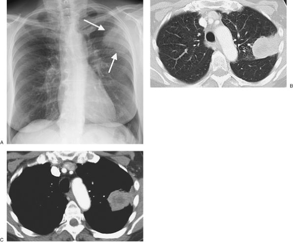 |
FIGURE 9-3. Subpleural squamous cell bronchogenic carcinoma. A: Posteroanterior (PA) chest radiograph of a 67-year-old woman shows a mass in the left upper hemithorax (arrows) that is contiguous with the pleural surface. B: CT with lung windowing shows the mass abutting the lateral pleural surface and major fissure. C:
CT with mediastinal windowing shows that the mass is contiguous with
the pleural surface. Centrally, the mass contains areas of low
attenuation, consistent with necrosis. |
Pleural Effusions
Pleural effusions develop
when there is excess pleural fluid produced, diminished resorption of
fluid from the pleural space, or both. The fluid can originate from the
pleura or be extrapleural in origin (Fig. 9-5).
Pleural effusions are categorized as either transudates or exudates.
The distinction is based on the specific gravity, protein, and lactate
dehydrogenase (LDH) content of the fluid, with transudates having a
specific gravity of 1.016 or less, a protein content of 3 g/dL or less,
a ratio of pleural fluid protein to serum protein of =0.5, an LDH ratio
(pleural fluid to serum) of =0.6, and an absolute pleural LDH level of
=200 IU/L (4). [AU: Fourth
sentence under subhead "Pleural Effusions": Are these last three
symbols (for ratios/levels) correct? They did not translate correctly,
so = (less than or equal to) was my best guess here. Please review
carefully and correct as needed.] Transudates develop primarily
as a result of changes in microvascular pressure and plasma oncotic
pressure, whereas exudates are caused by an altered pleural surface,
with an increase in permeability or a decrease in lymph flow. The list
of causes of pleural effusions is lengthy (Table 9-1),
but more than 90% of effusions result from heart failure, ascites,
pleuropulmonary infection, malignancy, or pulmonary embolism (5).
TABLE 9-1 COMMON CAUSES OF PLEURAL EFFUSION | |
|---|---|
|
P.141
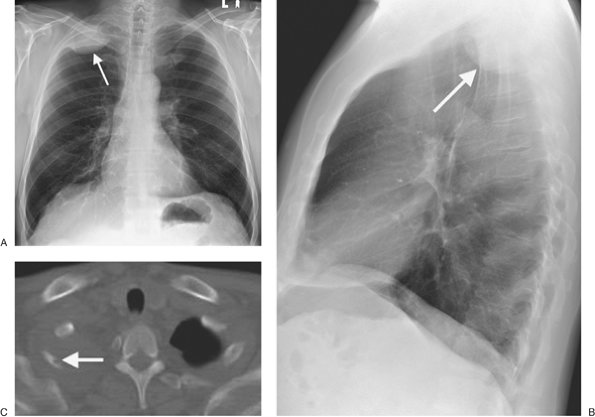 |
FIGURE 9-4. Pancoast tumor. PA (A) and lateral (B)
chest radiographs of a 61-year-old man with right shoulder pain and a
40–pack-year history of cigarette smoking shows a circumscribed mass (arrow) in the right apex. C: CT with bone windowing shows the mass filling the right lung apex and destruction of the right second rib (arrow). |
The appearance of an effusion depends on the patient's
position at the time of the radiologic examination and on the mobility
of the effusion. In an upright person, fluid collects mainly in the
lower pleural space, as long as it is freely mobile, creating a
homogeneous opacity with a curvilinear upper border that is sharply
marginated and concave to the lung (Fig. 9-6).
Fluid can collect in the fissures, creating a "pseudotumor" that
conforms to the edges of the fissures and resolves with clearing of
lung edema (Figs. 9-7 and 9-8). Occasionally, large quantities of pleural fluid accumulate in a "subpulmonic location"
rather than in the general pleural cavity. In this case, the upper edge of the fluid mimics the contour of the diaphragm on the chest radiograph, creating the appearance of an "elevated diaphragm," which usually peaks more laterally than normal. When this occurs on the left, the gastric air bubble and upper surface of the "hemidiaphragm" are separated more than usual. In supine patients, freely mobile fluid layers posteriorly, creating hazy, veillike opacification of the affected hemithorax with preserved vascular shadows. Depending on the size of the effusion, this can be a subtle finding; when bilateral, it may not be detected at all, especially when small, or it may be confused with pulmonary edema. Other findings of pleural effusions in supine patients include blunting of the costophrenic angle (although this is often a false-positive finding) (6), capping of the lung apex, thickening of the minor fissure, and widening of the paraspinal soft tissues. Lateral decubitus views can be useful in verifying pleural effusions when the supine examination is equivocal, and they can allow determination of whether pleural fluid is mobile or not. The lateral decubitus view is much more sensitive than the upright view for the detection of pleural effusions; it can demonstrate as little as 5 mL of pleural fluid (Fig. 9-9) (7).
P.142
P.143
rather than in the general pleural cavity. In this case, the upper edge of the fluid mimics the contour of the diaphragm on the chest radiograph, creating the appearance of an "elevated diaphragm," which usually peaks more laterally than normal. When this occurs on the left, the gastric air bubble and upper surface of the "hemidiaphragm" are separated more than usual. In supine patients, freely mobile fluid layers posteriorly, creating hazy, veillike opacification of the affected hemithorax with preserved vascular shadows. Depending on the size of the effusion, this can be a subtle finding; when bilateral, it may not be detected at all, especially when small, or it may be confused with pulmonary edema. Other findings of pleural effusions in supine patients include blunting of the costophrenic angle (although this is often a false-positive finding) (6), capping of the lung apex, thickening of the minor fissure, and widening of the paraspinal soft tissues. Lateral decubitus views can be useful in verifying pleural effusions when the supine examination is equivocal, and they can allow determination of whether pleural fluid is mobile or not. The lateral decubitus view is much more sensitive than the upright view for the detection of pleural effusions; it can demonstrate as little as 5 mL of pleural fluid (Fig. 9-9) (7).
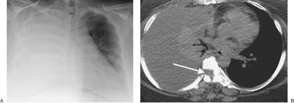 |
FIGURE 9-5. Cerebrospinal fluid leak into pleural space. A:
PA chest radiograph of a 42-year-old man who recently underwent partial
corpectomy of the thoracic spine at several levels shows complete
opacification of the right hemithorax and shift of the mediastinum to
the left. B: Non–contrast-enhanced CT
shows a large right pleural effusion, collapse of the right lung,
mediastinal shift to the left, findings of corpectomy, and continuity
of fluid from the spine into the pleural space (arrow). |
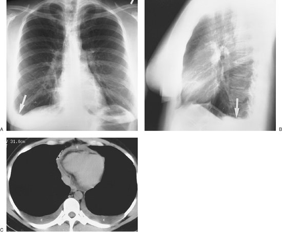 |
FIGURE 9-6. Pleural and pericardial effusions. A: PA chest radiograph of a woman with hypothyroidism shows blunting of the right costophrenic angle, producing a "meniscus" (arrow). B: Lateral chest radiograph shows blunting of both costophrenic angles posteriorly (arrow). C: CT shows bilateral pleural and pericardial effusions (E). |
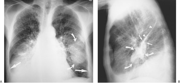 |
FIGURE 9-7. Pulmonary edema and pleural fluid pseudotumor. A:
PA chest radiograph shows enlargement of the cardiac silhouette,
interstitial pulmonary edema, and displacement of the inferolateral
lungs from the chest wall and diaphragm by pleural effusion (straight arrows). There is a hazy "mass" in the left middle and lower hemithorax (curved arrows). B: Lateral chest radiograph shows that the "mass" or "pseudotumor" (curved arrows) blends in with the left major fissure (straight arrows);
this is characteristic of pleural fluid within the fissure. The
superior aspect of the left major fissure is thickened as a result of
pleural fluid and subpleural edema (arrowheads). |
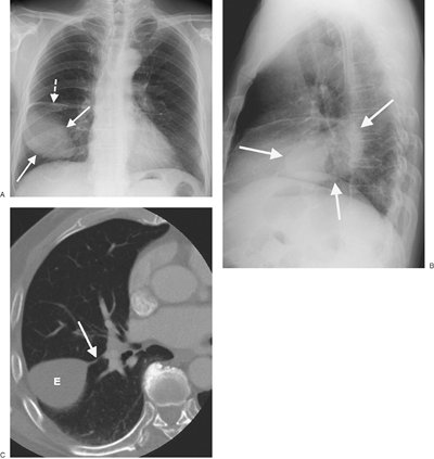 |
FIGURE 9-8. Pleural fluid pseudotumor. A: PA chest radiograph shows a circumscribed ovoid mass in the right lower hemithorax (solid arrows) and thickening of the minor fissure (dashed arrow). B: Lateral view shows that the mass (arrows) is oriented in the direction of and superimposed on the major fissure. C: CT (bone windowing) shows that the mass is of fluid attenuation, representing pleural effusion (E), and is contiguous with the thickened major fissure (arrow). |
CT can detect smaller amounts of pleural fluid than can chest radiography (8).
In addition, CT enables determination of whether fluid is loculated,
the extent and localization of loculated fluid for purposes of
drainage, assessment of pleural
morphology (irregular thickening and focal masses suggest malignancy), evaluation of underlying parenchymal disease, and differentiation between pleural and parenchymal disease (aided by the use of intravenous contrast material). The attenuation value of pleural fluid on CT enables detection of a hemothorax (Fig. 9-10), which has a higher attenuation value than simple fluid; occasionally, a fluid–fluid or hematocrit level can be seen (see Fig. 8-24). [AU: Did you intend to refer to Fig. ‘8-34’ here, rather than Fig. 8-24 (the latter shows a diaphragm rupture, whereas the former shows a hemothorax)?]
P.144
P.145
morphology (irregular thickening and focal masses suggest malignancy), evaluation of underlying parenchymal disease, and differentiation between pleural and parenchymal disease (aided by the use of intravenous contrast material). The attenuation value of pleural fluid on CT enables detection of a hemothorax (Fig. 9-10), which has a higher attenuation value than simple fluid; occasionally, a fluid–fluid or hematocrit level can be seen (see Fig. 8-24). [AU: Did you intend to refer to Fig. ‘8-34’ here, rather than Fig. 8-24 (the latter shows a diaphragm rupture, whereas the former shows a hemothorax)?]
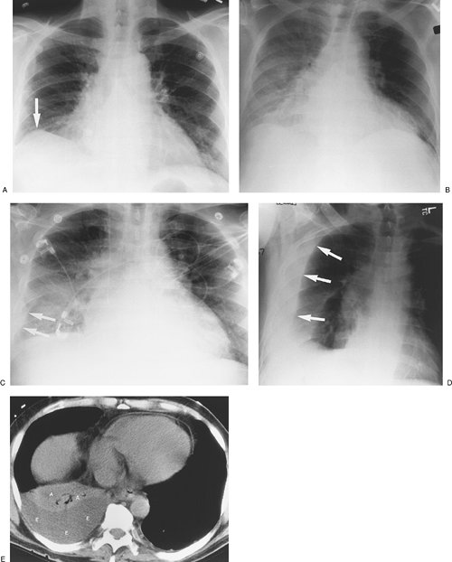 |
FIGURE 9-9. Positional appearances of pleural fluid on chest radiography and CT. A:
PA upright chest radiograph shows apparent elevation of the right
hemidiaphragm. The dome of the right hemidiaphragm appears to be
displaced laterally (arrow), a clue to the diagnosis of pleural fluid collecting in a "subpulmonic" location. B:
Anteroposterior (AP) supine chest radiograph of the same patient, 3
days later, shows hazy increased opacification of the right hemithorax
secondary to pleural fluid layering posteriorly within the pleural
space, now the most gravity-dependent portion of the pleural space. C: AP semi-upright chest radiograph of the same patient, 2 days after (B),
shows a combination of pleural fluid layering posteriorly, which
produces a hazy opacity in the mid and lower right hemithorax and
laterally (arrows). D: Right lateral decubitus chest radiograph of the same patient, taken on the same day as (A),
shows pleural fluid freely layering against the now gravity-dependent
lateral chest wall, from the costophrenic angle to the lung apex (arrows). E: CT of the same patient, performed on the same day as (B), shows a moderate- to large-sized right pleural fluid collection (E), with associated "passive" atelectasis of the right lower lobe (A). |
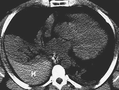 |
FIGURE 9-10. Hemothorax. CT shows high-attenuation blood (H) in the right pleural space. |
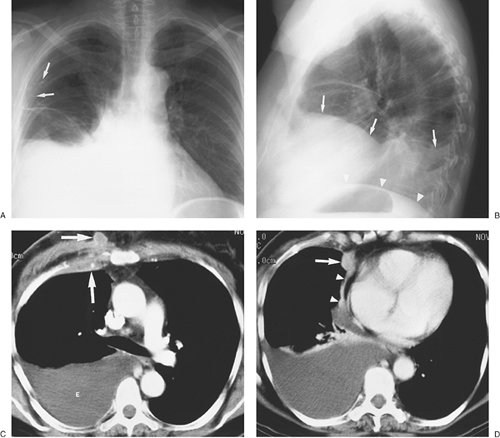 |
FIGURE 9-11. Malignant pleural effusion. A:
PA chest radiograph of a 62-year-old woman with metastatic breast
cancer who has had a right mastectomy and axillary node dissection
(note surgical clips in right axilla; arrows) shows apparent elevation of the right hemidiaphragm. B: Lateral chest radiograph also shows apparent elevation of the right hemidiaphragm (arrows). The left hemidiaphragm is easily identified (arrowheads), as it is just superior to the stomach bubble. C: CT shows a large right pleural effusion (E) and metastatic breast cancer infiltrating the right chest wall (arrows). D: CT at a level inferior to (C) shows a metastatic soft tissue mass to the mediastinal pleura (arrow) and thickening of the mediastinal pleura (arrowheads). |
When a large unilateral effusion is present (Fig. 9-11),
four causes should be considered: (i) infection (empyema); (ii) tumor
(primary bronchogenic carcinoma, mesothelioma, metastases, and
lymphoma); (iii) chylothorax (secondary to tumor, most notably
lymphoma, or ruptured thoracic duct); and (iv) hemorrhage (usually from
trauma, whether iatrogenic or otherwise) (Table 9-2).
Following drainage of a pneumothorax or pleural effusion, the
re-expanded lung may become acutely edematous. The edema usually
develops within 2 hours of re-expansion, can progress for 1 or 2 days,
and resolves within 5 to 7 days. Large pleural collections with
complete collapse
of the underlying lung, especially when long-standing, predispose to the development of re-expansion pulmonary edema (Fig. 9-12).
P.146
of the underlying lung, especially when long-standing, predispose to the development of re-expansion pulmonary edema (Fig. 9-12).
TABLE 9-2 CAUSES OF A LARGE UNILATERAL PLEURAL EFFUSION | |
|---|---|
|
Empyemais defined as pus in
the pleural cavity. The diagnosis is made when the pleural fluid is
obviously purulent, when organisms are identified in the fluid, or when
the fluid has an elevated white blood cell count. An empyema is assumed
to be present and drainage is indicated when there is associated
pneumonia and the fluid pH is below 7.0 or the fluid glucose level is
less than 40 mg/dL (9). The radiographic
appearance of empyema is that of pleural fluid, which is usually
unilateral but when bilateral is substantially greater in volume on the
infected side (Fig. 9-13). In contrast to
transudative pleural fluid collections, which typically have a smooth
margin that is concave to the lung, an empyema will often have a smooth
margin that is convex to the lung. Certain CT findings are suggestive
of empyema and other exudative effusions, including thickening and
enhancement of the parietal and visceral pleura (creating the “split
pleura sign”) after administration of intravenous contrast material (Figs. 9-14 and 9-15),
thickening of the extrapleural subcostal tissues, and increased
attenuation of the extrapleural fat. In some cases, empyema can be
difficult to distinguish from lung abscess. In general, there is a
sharply defined border between an empyema and the lung with
displacement and bowing of vessels and bronchi away from the empyema,
whereas abscesses lack a discrete boundary between the lesion and the
lung parenchyma. Empyemas are often elliptic and have a smooth inner
surface, whereas abscesses are more often round and have a relatively
thick wall. These features are not always reliable, however, and
occasionally it may be impossible to distinguish parenchymal from
pleural fluid collections (10).
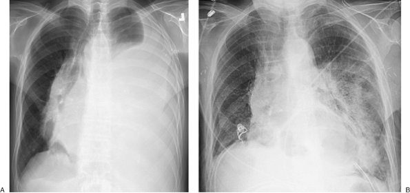 |
FIGURE 9-12. Re-expansion pulmonary edema. A:
PA chest radiograph of a 78-year-old woman with metastatic breast
cancer shows a large left pleural effusion associated with collapse of
the left lung and shift of the mediastinum to the right. These findings
suggest tension hydrothorax. B: PA chest
radiograph after placement of a left chest tube and adequate drainage
of pleural fluid shows re-expansion pulmonary edema on the left. |
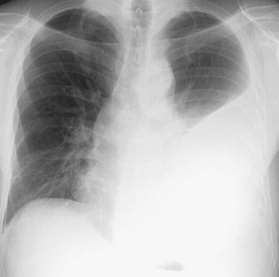 |
FIGURE 9-13. Tuberculous empyema.
PA chest radiograph shows a large left pleural effusion. A large
unilateral pleural effusion is worrisome for empyema, hemothorax,
malignancy, or chylothorax. |
P.147
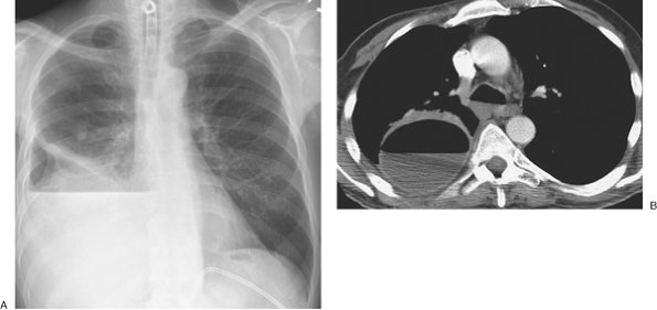 |
FIGURE 9-14. Empyema. A:
PA chest radiograph of a 60-year-old man with right lower lobe
pneumonia shows a large right hydropneumothorax with air–fluid level.
There is an incidental calcified granuloma in the right mid lung. B:
CT shows a round collection of air and fluid in the right pleural
space. The thickened and enhancing separated pleural layers create the
"split pleura" sign. Air within an empyema can be secondary to
thoracentesis, bronchopleural fistula, or, rarely, a gas-forming
organism. |
A chylothoraxcontains fluid
that is largely chyle (lymph of intestinal origin). Because chyle
usually contains suspended fat in the form of chylomicrons, chylothorax
fluid may be milky. Three main mechanisms account for chyle collections
in the pleural space: (i) leakage from a discrete rupture of the
thoracic duct or a large lymphatic vessel, (ii) a general oozing from
pleural lymphatics, and (iii) passage of chylous ascites through the
diaphragm (11). Approximately 50% of chylothoraces are of neoplastic origin, 25% are traumatic, and 15% are idiopathic (12). Lymphomas make up about 75% of the neoplastic lesions (13),
and chylothorax can be the initial feature of lymphomas. The CT
attenuation of chyle, despite its fat content, is usually
indistinguishable from that of other effusions because chyle is protein
rich.
Hemothoraxusually results
from trauma, either blunt or penetrating trauma to the chest or
iatrogenic trauma (such as with central venous catheter placement) (14).
A rapidly accumulating pleural fluid collection following trauma is
likely of arterial origin. High-pressure bleeding from systemic vessels
may be rapid and persistent, with the formation of a tension
hemothorax. CT of hemothorax may show areas of hyperdensity (15).
With clotting of the blood, loculation occurs; if undrained, a
hemothorax may eventually organize and cause pleural thickening
(fibrothorax).
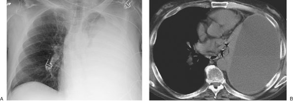 |
FIGURE 9-15. Empyema. A:
PA chest radiograph of a 55-year-old man shows a large left pleural
effusion, compression of the upper lung, and collapse of the lower
lung. B: CT shows an elongated ovoid collection of fluid in the left pleural space and collapse of the adjacent lung. |
Malignant pleural effusions are usually the result of metastases (95% of cases) (16), with bronchogenic cancer accounting for 36% of cases, breast cancer for 25% (Fig. 9-16), lymphoma for 10%, and ovarian and gastric carcinoma for 5% or fewer (Fig. 9-17) (17).
Although pleural effusion is often the major component of metastatic
disease to the pleura, other findings include pleural nodules or
extensive pleural thickening similar to that of mesothelioma. When the
pleural metastases are unilateral, the CT findings may be
indistinguishable from those of mesothelioma (18).
Malignant mesothelioma is a relatively rare primary
tumor of the pleura. Approximately 80% of these lesions occur in
individuals who have been exposed to asbestos (19).
The lifetime risk for the development of mesothelioma in asbestos
workers approaches 10%, and the average latency period is 35 years (20). Radiographic and CT findings include
nodular or irregular thickening of the visceral and parietal pleura, variable ipsilateral volume loss in the hemithorax, ipsilateral pleural effusion, involvement of the interlobar fissures and mediastinal pleural surfaces, and often fixation of the mediastinum (Figs. 9-18, 9-19, 9-20) (21). Approximately 18% of cases are associated with invasion of the chest wall (21).
P.148
nodular or irregular thickening of the visceral and parietal pleura, variable ipsilateral volume loss in the hemithorax, ipsilateral pleural effusion, involvement of the interlobar fissures and mediastinal pleural surfaces, and often fixation of the mediastinum (Figs. 9-18, 9-19, 9-20) (21). Approximately 18% of cases are associated with invasion of the chest wall (21).
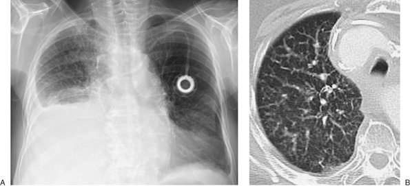 |
FIGURE 9-16. Malignant pleural effusion. A:
PA chest radiograph of an 83-year-old woman with metastatic right
breast cancer shows a large right pleural effusion and interstitial
lung disease. B: CT after drainage of
right pleural fluid shows nodular thickening of the vascular structures
and pulmonary septae on the right, characteristic of lymphangitic
carcinomatosis. |
Pneumothorax
Pneumothorax is defined as a collection of air in the pleural cavity and is divided into spontaneous and traumatic types (Table 9-3). A pneumothorax occurring without an obvious precipitating traumatic event or in a healthy individual is a primary spontaneous pneumothorax. This type of pneumothorax is strongly associated with smoking and tall asthenic men (22). Most patients are between 20 and 40 years of age, and the male-to-female ratio is approximately 5 to 1 (23).
The cause is nearly always the rupture of an apical pleural bulla.
Without treatment, the likelihood of another pneumothorax is about 40%,
and the chance of recurrence rises with each episode (23).
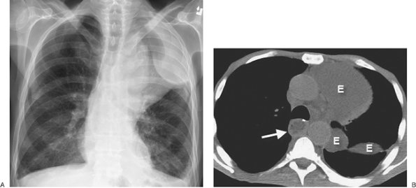 |
FIGURE 9-17. Malignant pleural effusion. A:
PA chest radiograph of a 57-year-old man with metastatic esophageal
carcinoma who had undergone esophagectomy and gastric pull-through
shows a lobulated opacity in the left upper hemithorax. B: CT shows loculated pleural fluid (E) extending into the fissure. There is fluid in the intrathoracic stomach (arrow). |
A pneumothorax developing without a precipitating traumatic event in a patient with predisposing lung disease is said to be a spontaneous secondary pneumothorax (Figs. 9-21 and 9-22).
Chronic obstructive pulmonary disease is the most common cause of
secondary spontaneous pneumothorax. About 0.5% of pneumothoraces are
associated with lung metastases, of which 89% are caused by sarcomas,
with osteogenic
sarcoma being the most common (24,25). Catamenial pneumothorax is an uncommon disorder that occurs in women, probably caused by air entering the peritoneal cavity by way of the genital tract during menses and proceeding to the pleural cavity through diaphragmatic fenestrations. Catamenial pneumothorax occurs only in relation to the menses, appearing 1 day before or up to 3 days after menses. The pneumothorax is usually small and most often right sided (87%) (26). Recurrence is a characteristic feature of catamenial pneumothorax, and it may be prevented by pregnancy or drugs that suppress ovulation.
P.149
P.150
sarcoma being the most common (24,25). Catamenial pneumothorax is an uncommon disorder that occurs in women, probably caused by air entering the peritoneal cavity by way of the genital tract during menses and proceeding to the pleural cavity through diaphragmatic fenestrations. Catamenial pneumothorax occurs only in relation to the menses, appearing 1 day before or up to 3 days after menses. The pneumothorax is usually small and most often right sided (87%) (26). Recurrence is a characteristic feature of catamenial pneumothorax, and it may be prevented by pregnancy or drugs that suppress ovulation.
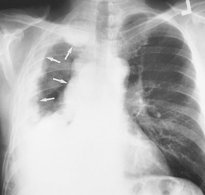 |
FIGURE 9-18. Malignant mesothelioma.
PA chest radiograph of a 53-year-old man shows right pleural
opacification with a lobulated contour that involves the entire pleural
surface (arrows) and is associated with a
"fixed mediastinum," meaning no shift right or left, and ipsilateral
loss of lung volume, characteristic of malignant mesothelioma. |
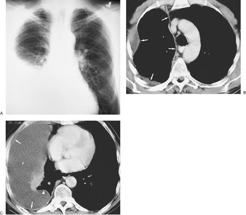 |
FIGURE 9-19. Malignant mesothelioma. A:
PA chest radiograph of a 69-year-old man who worked at a manufacturing
plant making brake linings and asbestos shingles shows a large
unilateral right pleural effusion. B: CT shows thickening of the entire pleural surface (arrows). C: CT scan at a level inferior to (B) shows a large right pleural effusion (E) and thickening and enhancement of the parietal (arrows) and visceral (arrowhead) pleura. |
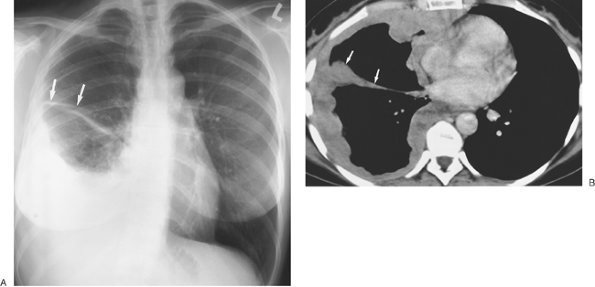 |
FIGURE 9-20. Malignant mesothelioma. A: PA chest radiograph of a 43-year-old man shows a large right pleural effusion with thickening of the minor fissure (arrows). B:
CT shows that the pleural effusion is of higher attenuation compared
with simple fluid (consistent with tumor within the pleural space);
wraps around the entire pleural surface, including the fissure (arrows); and has a lobulated contour. |
As with pleural effusion, the radiographic appearance of
pneumothorax depends on the radiographic projection, the patient's
position, and the presence or absence of pleural adhesion and
subsequent loculation. In the upright patient, air rises in the pleural
space and separates the lung from the chest wall, allowing the
visceral–pleural line to become visible as a thin curvilinear opacity
between vessel-containing lung and the avascular pneumothorax space.
The pleural line remains fairly parallel to the chest wall. Curvilinear
shadows projected over the lung apex that may mimic the visible
visceral–pleural line of a pneumothorax include vascular lines, tubes,
clothing, bedding, hair, scapulae, skin folds (Fig. 9-23),
and the walls of bullae and cavities. Cysts, bullae, and cavities
usually have inner margins that are concave to the chest wall rather
than convex. In the supine patient, the highest part of the chest
cavity lies anteriorly or anteromedially at the base near the
diaphragm, and free pleural air rises to this region (Fig. 9-24). If the pneumothorax is small or moderate in size, the lung is not separated from the chest wall laterally or at the apex and
therefore the pneumothorax may not be appreciated. Signs of pneumothorax in a supine patient are listed in Table 9-4 (27).
P.151
P.152
therefore the pneumothorax may not be appreciated. Signs of pneumothorax in a supine patient are listed in Table 9-4 (27).
TABLE 9-3 CAUSES OF PNEUMOTHORAX IN ADULTS | |
|---|---|
|
 |
FIGURE 9-21. Spontaneous secondary pneumothorax.
PA chest radiograph of an 18-year-old man with cystic fibrosis shows a
large right hydropneumothorax and severe bilateral cystic
bronchiectasis. |
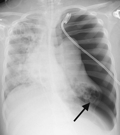 |
FIGURE 9-22. Spontaneous secondary pneumothorax.
AP chest radiograph of a 3-year-old boy with respiratory syncytial
virus pneumonia shows a large left pneumothorax with shift of the
mediastinum to the right. The findings suggest a tension pneumothorax.
Numerous cystic lesions, consistent with pneumatoceles, are seen in the
collapsed left lung (arrow). |
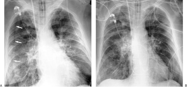 |
FIGURE 9-23. Skin fold mimicking pneumothorax. A: AP supine chest radiograph shows opacification of the right medial lung outlined by a sharp edge (skin fold; arrows). Note that the lung peripheral to this edge is not hyperlucent, a clue that there is no pneumothorax. B:
AP upright chest radiograph obtained 1 hour later no longer shows the
skin fold. Redundant skin can result in skin folds on the chest
radiograph, especially when the patient is supine. Changing patient
positioning is often useful in differentiating a skin fold from a
pneumothorax. |
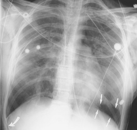 |
FIGURE 9-24. Pneumothorax in a supine patient. AP supine chest radiograph shows the "deep sulcus" sign of pneumothorax on the right (curved arrow) and a basilar pneumothorax on the left (straight arrows). |
TABLE 9-4 RADIOGRAPHIC SIGNS OF PNEUMOTHORAX IN THE SUPINE PATIENT | |
|---|---|
|
A large tension pneumothorax can be a life-threatening
situation requiring rapid decompression. Radiologic signs of tension
pneumothorax include contralateral displacement of the mediastinum,
inferior displacement of the diaphragm, hyperlucent hemithorax, and
ipsilateral collapse of the lung (Fig. 9-25).
Localized Pleural Tumors
Focal pleural tumors include
localized fibrous tumors of pleura, lipomas, liposarcomas, and invasion
of the pleura by adjacent bronchogenic carcinoma. Malignant
mesothelioma and metastases may cause focal abnormalities (Fig. 9-26)
but are most commonly associated with extensive involvement of the
pleura, as previously discussed. Localized fibrous tumor of the pleura
is either benign (60%) or malignant (40%) and has a relatively good
prognosis when surgically excised (28). It
occurs in all age groups, most commonly in patients older than 50, and
it is not related to asbestos exposure. Forty percent of these tumors
are attached to the pleura by a pedicle, and they may range from 1 to
39 cm in diameter. Calcification is present in 5% of cases. The
radiographic appearance is a mass with a smooth, sharply delineated
contour with tapering margins that forms obtuse angles with the chest
wall or mediastinum and displaces the adjacent lung parenchyma (Fig. 9-27). Pedunculated tumors may be mobile and change their position with respiration or posture.
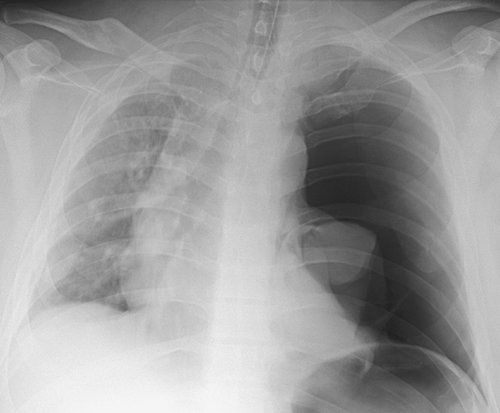 |
FIGURE 9-25. Tension pneumothorax.
AP supine chest radiograph of a 35-year-old man involved in a motor
vehicle crash shows a large left pneumothorax, collapse of the left
lung, and shift of the mediastinum to the right. The findings suggest a
tension pneumothorax, which requires immediate decompression. |
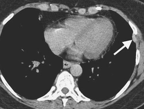 |
FIGURE 9-26. Malignant mesothelioma. CT scan of a 59-year-old man shows a solitary focal enhancing left pleural soft tissue lesion (arrow). At surgery, multiple additional lesions were seen studding the pleural surface. |
Pleural lipomas are rare tumors usually found
incidentally on the chest radiograph. On CT, a pleural lipoma appears
as a well-defined mass of homogeneous fat attenuation having obtuse
angles with the chest wall and displacing the adjacent pulmonary
parenchyma (Fig. 9-28) (29).
When the tumor is heterogeneous and has attenuation values greater than
about 50 Hounsfield units, a liposarcoma should be suspected (Fig. 9-29) (30).
Diffuse Pleural Fibrosis (Fibrothorax)
Fibrothorax (fibrous
obliteration of the pleural space) may develop as a result of organized
hemorrhagic effusions, tuberculous effusions, other types of empyema,
and extensive benign asbestos-related pleural disease. Asbestos-related
diffuse pleural thickening is much less common than discrete pleural
plaques and involves the visceral rather than the parietal pleura (31).
Evidence of underlying parenchymal disease is usually seen in patients
with prior tuberculosis or other empyema. Extensive calcification of
the fibrothorax favors previous tuberculosis, empyema, hemothorax or
trauma (Fig. 9-30) and is seldom seen with asbestos-related diffuse pleural thickening (32).
Hemorrhagic effusion, tuberculosis, and other causes of empyema usually
lead to unilateral pleural abnormalities, whereas benign asbestos
pleurisy usually leads to bilateral pleural abnormalities, whether
diffuse pleural thickening or pleural plaques. Involvement of the
mediastinal pleura is more common with mesothelioma or other
malignancies than with benign fibrothorax (18).
Pleural Plaques
Pleural plaques are
circumscribed collections of dense collagenous connective tissue, which
may or may not be calcified, and represent the most common
manifestation of asbestos exposure (31). The latency period between exposure to asbestos and development of pleural plaques is approximately
15 years. The plaques involve mainly the posterior and anterolateral aspects of the pleura, following the contours of the posterolateral seventh to 10th ribs, and the domes of the hemidiaphragms, and spare the lung apices and costophrenic angles. They almost always involve only the parietal pleura but occasionally may be seen in the visceral pleura in the interlobar fissures and sometimes involve the pericardium (Fig. 9-31). On chest radiographs, pleural plaques are unilateral in approximately 25% of cases (33), although more plaques are detected on CT than chest radiography. Pleural plaques are not premalignant, but detection of them is important for three main reasons: (i) in patients with associated interstitial lung disease, the presence of pleural plaques, in the appropriate clinical setting, strongly suggests the diagnosis of asbestosis; (ii) they are virtually pathognomonic of asbestos exposure and should prompt an occupational history; and (iii) they may encourage a patient to stop smoking, because there is a synergistic interaction between asbestos exposure and smoking in the development of lung cancer. Asbestos-related pleural disease has five manifestations: (i) pleural plaque with or without calcification, (ii) asbestos-related pleural effusion, (iii) diffuse pleural thickening, (iv) rounded atelectasis, and (v) mesothelioma.
P.153
P.154
P.155
15 years. The plaques involve mainly the posterior and anterolateral aspects of the pleura, following the contours of the posterolateral seventh to 10th ribs, and the domes of the hemidiaphragms, and spare the lung apices and costophrenic angles. They almost always involve only the parietal pleura but occasionally may be seen in the visceral pleura in the interlobar fissures and sometimes involve the pericardium (Fig. 9-31). On chest radiographs, pleural plaques are unilateral in approximately 25% of cases (33), although more plaques are detected on CT than chest radiography. Pleural plaques are not premalignant, but detection of them is important for three main reasons: (i) in patients with associated interstitial lung disease, the presence of pleural plaques, in the appropriate clinical setting, strongly suggests the diagnosis of asbestosis; (ii) they are virtually pathognomonic of asbestos exposure and should prompt an occupational history; and (iii) they may encourage a patient to stop smoking, because there is a synergistic interaction between asbestos exposure and smoking in the development of lung cancer. Asbestos-related pleural disease has five manifestations: (i) pleural plaque with or without calcification, (ii) asbestos-related pleural effusion, (iii) diffuse pleural thickening, (iv) rounded atelectasis, and (v) mesothelioma.
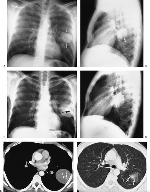 |
FIGURE 9-27. Benign localized fibrous tumor of the pleura. A: PA chest radiograph of a 32-year-old asymptomatic woman shows a well-circumscribed round mass in the left middle lung (arrows). B: Lateral view shows that the mass is positioned adjacent to the left major fissure (arrows). C:
PA chest radiograph obtained 4 years later shows that the mass has
increased in size. Faint calcification is now visible within the mass (arrow). D: Lateral view obtained at the same time as (C). E: CT shows that the mass is homogeneous, abuts the lateral pleural surface, and has coarse calcifications (arrows). F: CT with lung windowing shows that the top of the tumor abuts the major fissure (arrows). |
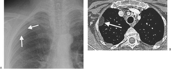 |
FIGURE 9-28. Pleural lipoma. A: PA chest radiograph coned to the right upper lobe shows a circumscribed mass (arrows) contiguous with the right lateral chest wall. B: CT shows that the mass (arrow) is of homogeneous fat attenuation (F). |
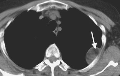 |
FIGURE 9-29. Benign pleural myelolipoma.
CT of a 66-year-old woman shows a circumscribed mass abutting the left
lateral chest wall. Although the mass contains central fat (arrow),
the attenuation of the mass is heterogeneous, and a malignancy, such as
liposarcoma, cannot be excluded. The mass was excised and found to be
benign. |
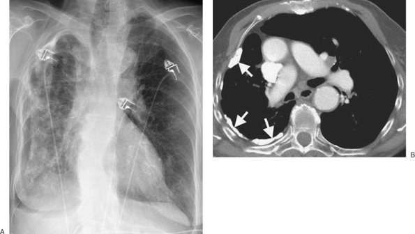 |
FIGURE 9-30. Old tuberculous empyema. A: PA chest radiograph shows dense calcification throughout the right hemithorax. B:
Chest radiograph with dual energy, to emphasize the bones and calcium,
optimally shows the long linear calcifications in the right hemithorax.
C: CT shows dense pleural calcifications (arrows) involving only the right hemithorax, associated with loss of lung volume. [AU/PRODUCTION:
Note that 9-30B should be a chest radiograph; what is labeled as 9-30B
appears to in fact be 9-30C, and 9-30B is missing.] |
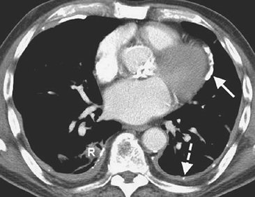 |
FIGURE 9-31. Calcified pleural and pericardial plaques. CT of an 82-year-old man with prior asbestos exposure shows calcified pericardial (solid arrow) and pleural (dashed arrow) plaques, in addition to diffuse pleural thickening and rounded atelectasis (R). |
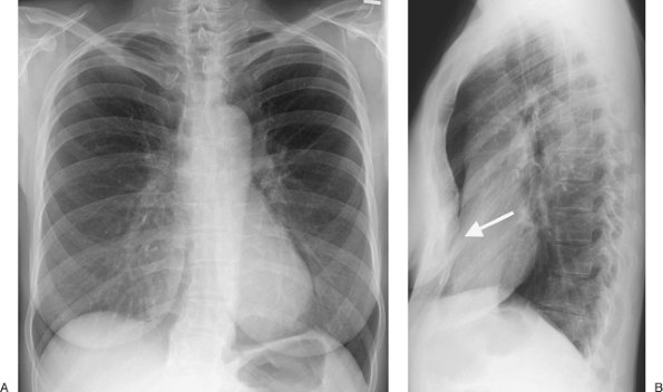 |
FIGURE 9-32. Pectus excavatum. A:
PA chest radiograph of a 60-year-old man shows blurring of the right
heart border and displacement of the mediastinum to the left. B: Lateral view shows posterior depression of the sternum (arrow). |
Chest Wall
The thoracic contents are bounded by the chest wall,
which consists of skin, subcutaneous tissues, muscles, clavicles,
scapulae, ribs, sternum, and spine. Deformities and normal variants of
chest wall anatomy are relatively common. Accessory cervical ribs,
arising from the seventh cervical vertebra as enlarged costal elements,
occur in approximately 0.5% to 1.5% of the population and may lead to
thoracic outlet syndrome (34). The most common
sternal deformity is pectus excavatum, which is easily appreciated on a
lateral chest radiograph as posterior depression of the sternum. On a
frontal radiograph, it may cause blurring of the right heart border and
displacement of the heart to the left, mimicking a right middle lobe
process (Fig. 9-32).
En face, chest wall lesions characteristically appear as
homogeneous, often partly rounded opacities with a sharp medial edge
and an ill-defined lateral margin. Tangentially, chest wall and
localized pleural lesions are convex to the lung and sharply
marginated, since they are covered on the lung aspect by pleura (Fig. 9-33).
Chest wall masses usually have an obtuse angle of contact with the
chest wall. On chest radiographs, localized lesions of the chest wall
frequently cannot be distinguished from localized pleural lesions
unless there is rib remodeling or destruction. Rib involvement is
characteristic of a chest wall lesion and, except for the occasional
invasive neoplastic or infective lesion (actinomycosis, tuberculosis),
is rarely seen with pleural or lung processes. Masses involving the
chest wall include infectious and benign and malignant neoplastic
causes (Figs. 9-34, 9-35, 9-36, 9-37, 9-38, 9-39, 9-40) (Table 9-5).
P.156
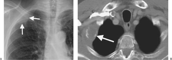 |
FIGURE 9-33. Lymphoma with rib involvement. A: PA chest radiograph shows a circumscribed mass (arrows) that is contiguous with the upper right lateral chest wall. B: CT shows the mass (arrow) and destruction of the right second rib. |
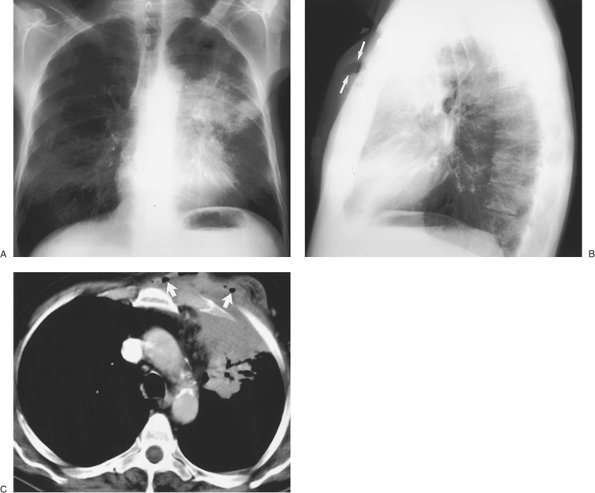 |
FIGURE 9-34. Actinomycosis of the chest wall. A:
PA chest radiograph of a 58-year-old man with a 1-month history of
shortness of breath and poor oral hygiene shows airspace disease in the
left upper lobe. B: Lateral view shows
extension of the left upper lobe pneumonia into the anterior chest
wall. There is air within the swollen soft tissues of the chest wall (arrows). C:
CT shows extension of the left upper lobe pneumonia into the anterior
chest wall, along with numerous bubbles of air within the chest wall (arrows). |
P.157
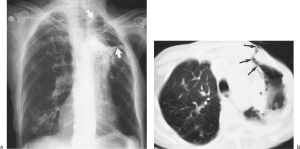 |
FIGURE 9-35. Tuberculous pneumonia with broncho-pleural-cutaneous fistula. A:
AP upright chest radiograph of an 83-year-old woman with a history of
left mastectomy and cobalt radiotherapy 30 years prior, along with a
remote history of positive skin test for tuberculosis that was treated
with appropriate drug therapy. Now, with a draining left chest wall
wound at the surgical scar site, the patient's chest radiograph shows a
cavitary mass in the left upper lobe (arrows) and scarring in the right upper lobe (unchanged compared with prior chest radiographs). B: CT shows air from the cavity communicating with the pleura and skin of the anterior chest wall (arrows), so-called empyema necessitatis. Analysis of the fluid draining from the chest wall revealed Mycobacterium tuberculosis.
The patient was predisposed to the development of reactivation
tuberculosis because she had recently been taking high doses of
steroids to treat polymyalgia rheumatica. |
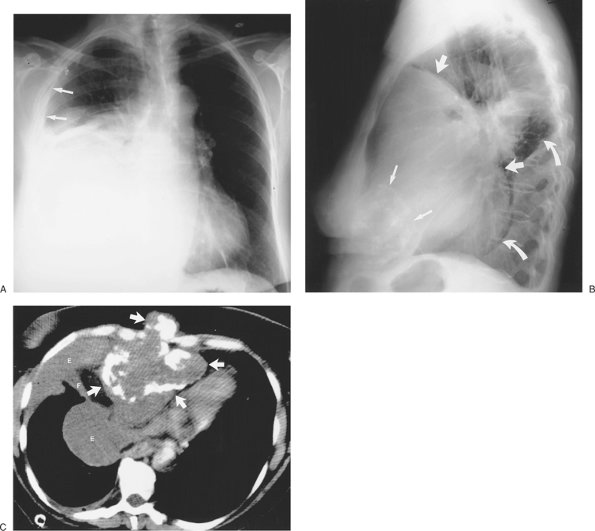 |
FIGURE 9-36. Chondrosarcoma of the sternum. A:
PA chest radiograph of a 67-year-old woman shows a large area of
opacification in the right lower hemithorax, a right pleural effusion (arrows), and shift of the mediastinum to the left. B: Lateral view shows amorphous calcifications overlying the anterior heart (smaller arrows), a large mass within the anterior mediastinum (larger arrows), and a pleural effusion (curved arrows). C: CT, with patient prone, shows a calcified mass arising from the sternum (arrows) and a loculated right malignant pleural effusion (E) extending into the major fissure (F). Differential diagnosis included malignant teratoma, malignant thymoma, and osteogenic sarcoma. |
P.158
 |
FIGURE 9-37. Sternal metastases.
CT of a 64-year-old woman with metastatic endometrial carcinoma shows
complete destruction of the sternum by a large soft tissue mass with
punctate and curvilinear calcifications. |
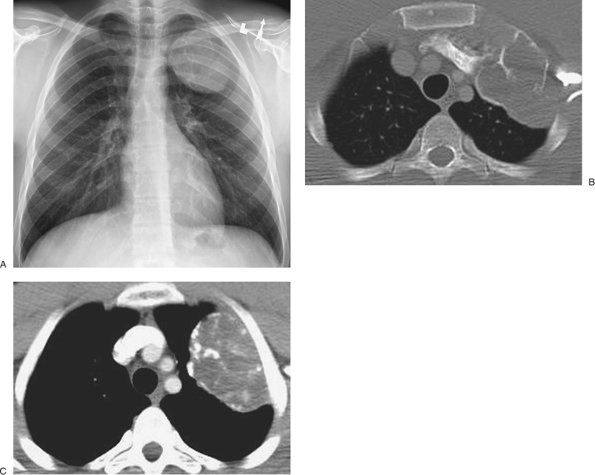 |
FIGURE 9-38. Aneurysmal bone cyst of the left chest wall. A:
PA chest radiograph of a 19-year-old asymptomatic man with a history of
Hodgkin disease treated with chemotherapy and radiation shows a
circumscribed mass projecting over the left upper lobe and contiguous
with the left upper chest wall. B: CT with bone windowing shows that the destructive mass arises from the left upper ribs. C: CT at a level inferior to (B)
shows that the mass contains areas of dense calcification. The
appearance of this new mass on routine chest radiography was suspicious
for a radiation-induced sarcoma. |
P.159
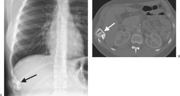 |
FIGURE 9-39. Benign rib osteochondroma. A: PA chest radiograph of a 20-year-old man shows a calcified mass arising form a lower right lateral rib (arrow). B:
CT shows continuity of the cortex and marrow of the osteochondroma with
that of the host rib, as well as characteristic extension of the
osteochondroma on a stalk with a cauliflowerlike head. |
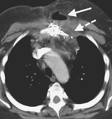 |
FIGURE 9-40. Sternal wound infection. CT scan of a 47-year-old man several weeks after coronary artery bypass grafting shows an air–fluid level (solid arrow) and a focal fluid collection (dashed arrow)
in the presternal soft tissues and abnormal areas of high attenuation
in the retrosternal area. Bacterial infection was confirmed during
surgical debridement. |
TABLE 9-5 MASSES INVOLVING THE CHEST WALL | |||
|---|---|---|---|
|
P.160
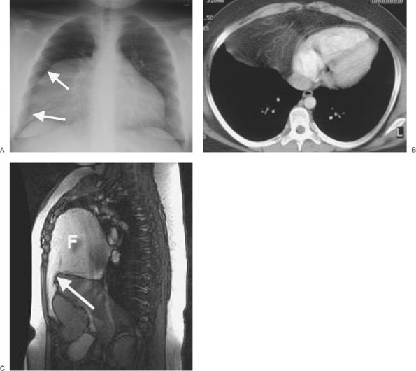 |
FIGURE 9-41. Foramen of Morgagni hernia. A: PA chest radiograph of an 11-year-old girl shows an abnormal right mediastinal contour (arrows) and loss of the normal right heart border. B: CT shows fat and prominent omental vascular structures anterior to the heart. C: Sagittal T1-weighted magnetic resonance image shows a defect in the anterior diaphragm (arrow) and herniation of high-signal fat (F) into the anterior chest. |
Diaphragm
The diaphragm consists of a large, dome-shaped central
tendon with a sheet of striated muscle radiating from the central
tendon to attach to the seventh through 12th ribs and to the
xiphisternum (35). The two crura arise from the
upper three lumbar vertebrae and arch upward and forward to form the
margins of the aortic hiatus and esophageal hiatus. The diaphragm has a
smooth dome shape in most individuals, but a scalloped outline is also
common. Radiographically, in most people, the right hemidiaphragm is
1.5 to 2.5 cm higher than the left, but the two hemidiaphragms are at
the same level in about 9% of the population, and the left
hemidiaphragm is higher than the right (by less than 1 cm) in about 3%
of the population (36). Incomplete muscularization, known as eventration,
is common and frequently involves the anteromedial portion of the
hemidiaphragm (usually the right, but occasionally affecting the left),
producing a smooth hump on the contour of the diaphragm.
Congenital hernias of the diaphragm are common. Ninety
percent of these are Bochdalek hernias, which arise posterolaterally
because of failure of the costal and vertebral portions of the
diaphragm to fuse. They are more common on the left than the right.
Small incidental Bochdalek hernias are seen frequently on CT, often
with no more than a small amount of retroperitoneal fat herniating
through the defect. Morgagni hernias, most commonly seen on the right,
arise anteromedially from failure of the sternal and costal portions of
the diaphragm to fuse (Fig. 9-41).
When unilateral elevation of the diaphragm is seen on chest radiography, five diagnostic possibilities should be considered (Table 9-6) (Figs. 9-42 and 9-43).
First, however, it is important to determine the chronicity of the
elevation, since stability for 2 or more years on chest radiography
makes a malignant cause highly unlikely and makes acute processes such
as lung collapse and subdiaphragmatic abscess less likely. Subpulmonic
effusion was discussed earlier in this chapter. Diaphragmatic rupture
was discussed in Chapter 8, and atelectasis is discussed in Chapter 11.
The phrenic nerves, which arise from the third through
fifth cervical nerves, supply the diaphragm (“C3, 4, 5 keep the
diaphragm alive”). The right phrenic nerve descends at the right side
of the superior vena cava and right atrium, in front of the
root of the right lung, between the pericardium and mediastinal pleura. The left phrenic nerve descends between the left subclavian and left common carotid arteries, lateral to the vagus nerve and the arch of the aorta. It passes in front of the root of the left lung between the mediastinal pleura and the pericardium, and its branches pierce the diaphragm immediately to the left of the pericardium. Tumor invading the phrenic nerve on either side can result in elevation of the ipsilateral diaphragm and should always be considered as the cause of an abnormally elevated diaphragm in patients over age 35 unless proven to be stable for 2 years or more on prior chest radiographs (Fig. 9-44). The chest radiograph may not show the tumor itself, and occasionally the elevated diaphragm may be the only radiographic clue to an underlying neoplasm.
P.161
P.162
root of the right lung, between the pericardium and mediastinal pleura. The left phrenic nerve descends between the left subclavian and left common carotid arteries, lateral to the vagus nerve and the arch of the aorta. It passes in front of the root of the left lung between the mediastinal pleura and the pericardium, and its branches pierce the diaphragm immediately to the left of the pericardium. Tumor invading the phrenic nerve on either side can result in elevation of the ipsilateral diaphragm and should always be considered as the cause of an abnormally elevated diaphragm in patients over age 35 unless proven to be stable for 2 years or more on prior chest radiographs (Fig. 9-44). The chest radiograph may not show the tumor itself, and occasionally the elevated diaphragm may be the only radiographic clue to an underlying neoplasm.
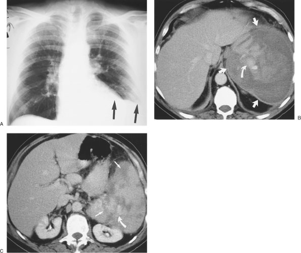 |
FIGURE 9-42. Elevated diaphragm secondary to subdiaphragmatic splenic hematoma. A:
AP upright chest radiograph of a 68-year-old man with left flank pain
and chronic lymphocytic lymphoma shows elevation of the left
hemidiaphragm (arrows). B: CT shows a large subcapsular splenic hematoma (straight arrows). The high-attenuation material within the hematoma represents acute bleeding (curved arrow). C: CT at a level inferior to (B) shows multiple low-attenuation areas within the spleen from spontaneous splenic rupture (straight arrows) and high-attenuation material from acute bleeding (curved arrow). |
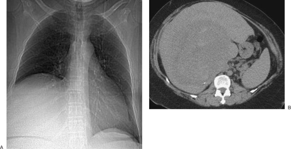 |
FIGURE 9-43. Elevated diaphragm secondary to hepatic hemangioma. A: CT scout image shows elevation of the right hemidiaphragm. B: Axial CT shows a large hepatic mass. |
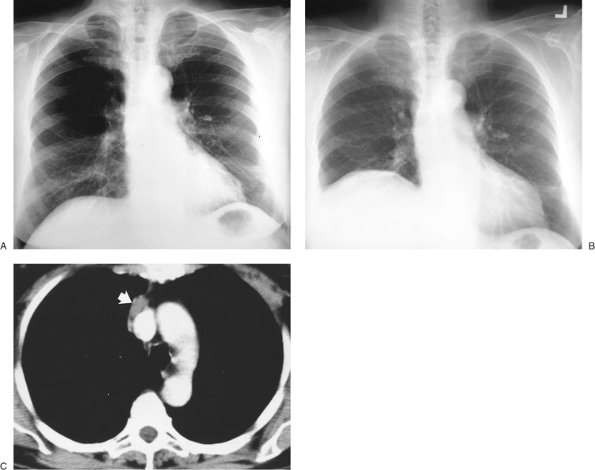 |
FIGURE 9-44. Bronchogenic carcinoma invading the phrenic nerve. A: PA chest radiograph of a 74-year-old woman shows normal positioning of the hemidiaphragms. B: PA chest radiograph obtained 1 year later shows elevation of the right hemidiaphragm and no evidence of mediastinal mass. C: CT shows a homogeneous soft tissue mass adjacent to the superior vena cava (arrows),
which proved to be a bronchogenic adenocarcinoma invading the right
phrenic nerve. This case illustrates the significance of new
diaphragmatic elevation in an adult, even when no mediastinal mass is
seen on the chest radiograph. |
TABLE 9-6 UNILATERAL ELEVATED DIAPHRAGM | |
|---|---|
|
References
1. Black LF. The pleural space and pleural fluid. Mayo Clin Proc. 1972;47:493–506.
2. Im JG, Webb WR, Rosen A, Gamsu G. Costal pleura: appearances at high-resolution CT. Radiology. 1989;171:125–131.
3. Groskin SA. Radiologic–pathologic correlations. In: Groskin SA, ed. Heitzman's The Lung. St. Louis, MO: Mosby; 1993:575–609.
4. Light RW. Pleural Diseases. Philadelphia: Lea & Febiger; 1983.
5. Jay SJ. Diagnostic procedures for pleural disease. Clin Chest Med. 1985;6:33–48.
6. Ruskin JA, Gurney JW, Thorsen MK, et al. Detection of pleural effusions on supine chest radiographs. AJR Am J Roentgenol. 1987;148:681–683.
7. Moskowitz H, Platt RT, Schacher R, Mellins H. Roentgen visualization of minute pleural effusion. Radiology. 1973;109:33–35.
8. McLoud TC, Flower CDR. Imaging the pleura: sonography, CT, and MR imaging. AJR Am J Roentgenol. 1991;156:1145–1153.
9. Light RW. Diseases of the pleura, mediastinum, chest wall, and diaphragm. In: George RB, Light RW, Matthay MA, et al, eds. Chest Medicine. Baltimore: Williams & Wilkins; 1990:318–412.
10. Naidich DP, Zerhouni EA, Siegelman SS. Pleura and chest wall. In: Naidich DP, Zerhouni EA, Siegelman SS, eds. Computed Tomography and Magnetic Resonance of the Thorax. 2nd ed. New York: Raven Press; 1991:407–471.
11. Nix JT, Albert M, Dugas JE, et al. Chylothorax and chylous ascites: a study of 302 selected cases. Am J Gastroenterol. 1957;28:40–53.
P.163
12. Sassoon CS, Light RW. Chylothorax and pseudochylothorax. Clin Chest Med. 1985;6:163–171.
13. MacFarlane JR, Holman CW. Chylothorax. Am Rev Respir Dis. 1972;105:287–291.
14. Groskin SA. Selected topics in chest trauma. Radiology. 1992;183:605–617.
15. Wolverson
MK, Crepps LF, Sundaram M, et al. Hyperdensity of recurrent hemorrhage
at body computed tomography: incidence and morphologic variation. Radiology. 1983;148:779–784.
16. Godwin JD, ed. Computed Tomography of the Chest. Philadelphia: Lippincott; 1984:130–137.
17. Sahn SA. Malignant pleural effusion. In: Fishman AP, ed. Pulmonary Diseases and Disorders. 2nd ed. New York: McGraw-Hill; 1988:2159–2169.
18. Leung AN, Müller NL, Miller RR. CT in differential diagnosis of diffuse pleural disease. AJR Am J Roentgenol. 1990;154:487–492.
19. Mossman BT, Gee JBL. Asbestos-related diseases. N Engl J Med. 1989;320:1721–1730.
20. Antman KH. Clinical presentation and natural history of benign and malignant mesothelioma. Semin Oncol. 1981;8:313–320.
21. Kawashima A, Libshitz HI. Malignant pleural mesothelioma: CT manifestations in 50 cases. AJR Am J Roentgenol. 1990;155:965–969.
22. Jansveld CAF, Dijkman JH. Primary spontaneous pneumothorax and smoking. Br Med J. 1975;4:559–560.
23. Killen DA, Gobbel WG. Spontaneous Pneumothorax. Boston: Little, Brown; 1968.
24. Dines DE, Cortese DA, Brennan MD, et al. Malignant pulmonary neoplasms predisposing to spontaneous pneumothorax. Mayo Clin Proc. 1973;48:541–544.
25. Janetos GP, Ochsner SF. Bilateral pneumothorax in metastatic osteogenic sarcoma. Am Rev Respir Dis. 1963;88:73–76.
26. Carter EJ, Ettensohn DB. Catamenial pneumothorax. Chest. 1990;98:713–716.
27. Tocino IM, Miller MH, Fairfax WR. Distribution of pneumothorax in the supine and semirecumbent critically ill adult. AJR Am J Roentgenol. 1985;144:901–905.
28. England
DM, Hochholzer L, McCarthy MJ. Localized benign and malignant fibrous
tumors of the pleura: a clinicopathologic review of 223 cases. Am J Surg Pathol. 1989;13:640–658.
29. Buxton RC, Tan CS, Khine NM, et al. Atypical transmural thoracic lipoma: CT diagnosis. J Comput Assist Tomogr. 1988;12:196–198.
30. Munk PL, Müller NL. Pleural liposarcoma: CT diagnosis. J Comput Assist Tomogr. 1988;12:709–710.
31. Schwartz DA. New developments in asbestos-related pleural disease. Chest. 1991;99:191–198.
32. Friedman
AC, Fiel SB, Radecki PD, et al. Computed tomography of benign pleural
and pulmonary parenchymal abnormalities related to asbestos exposure. Semin Ultrasound CT MR. 1990;11:393–408.
33. Withers BF, Ducatman AM, Yang WN. Roentgenographic evidence for predominant left-sided location of unilateral pleural plaques. Chest. 1984;95: 1262–1264.
34. Kurihara Y, Yakushiji YK, Matsumoto J, et al. The ribs: anatomic and radiologic considerations. RadioGraphics. 1999;19:105–119, 151–152.
35. Heitzman ER. The diaphragm: radiologic correlations with anatomy and pathology. Clin Radiol. 1990;42:15–19.
36. Felson B. Chest Roentgenology. Philadelphia: WB Saunders; 1973.